An overview of what we do at Victory Animal Hospital
我們在勝利動物醫院所做的工作概述
The knee is intrinsically supported by multiple ligaments, the main being the cruciate ligaments. There are 2 of these ligaments and they stabilize the femur and tibia relative to flexion and extension of the knee.
膝關節本質上由多條韌帶支撐,其中最主要的是十字韌帶。十字韌帶前後共有 2 條,在膝關節屈伸時穩定股骨和脛骨。
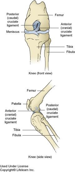
The cranial cruciate ligament is the most susceptible to injury and thus rupturing. When ruptured the vet will note a positive “drawer sign” or a “tibial thrust” on a compression test. This occurs most commonly in athletic large breed dogs but can occur in any size dog or cat.
前十字韌帶最容易受傷,因此亦最容易斷裂。一旦斷裂,獸醫會透過臨床檢查推測韌帶受損的程度。這種情況最常見於運動型大型犬,但也可能發生在任何體型的犬或貓身上。
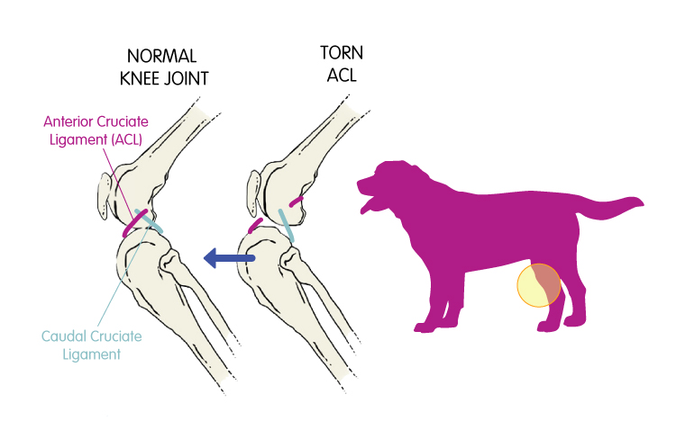
When examining patients with suspect CCL rupture the patients are often painful and guard the knee by tensing their muscles, thus sedation is often required. While sedated, radiographs can be taken and tests performed to confirm the diagnosis of CCL rupture.
在對疑似前十字韌帶斷裂的犬隻進行檢查時,他們通常會感到疼痛,並透過繃緊肌肉來保護膝蓋,因此檢查程序通常需要使用鎮靜劑。在鎮靜狀態下,可以拍攝X光片並進行檢查,以確診是否為前十字韌帶斷裂。
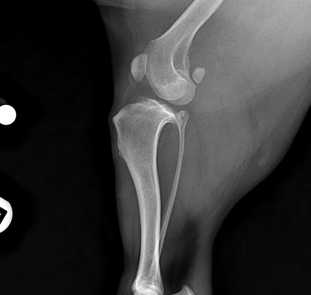
繼前十字韌帶斷裂後而導致的關節發炎–注意關節膨脹、脂肪墊消失、骨刺形成、軟組織腫脹和筋膜平面塌陷
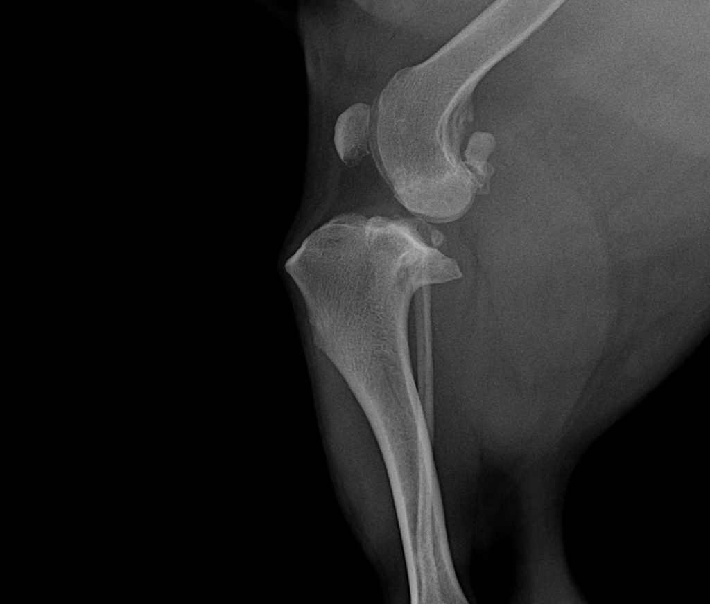
對側膝關節的 X 光片對比–看看大家能否發現X光中的不同
Once confirmed, treatments can be considered based on the degree of injury, Partial tears can be cage rested and platelet-rich plasma considered BUT Full ruptures will require surgery, unless the patient cannot tolerate the general anaesthesia due to underlying medical condition. Surgery is recommended due to the high rate of osteo-arthritic changes that occur in an unstable knee. The arthritis causes pain which reduces use of the limb, causing muscle wasting which further exacerbates the instability and progression of arthritis, creating a circle of pathology and deterioration
一旦確診,可根據損傷程度選擇治療方法,部分撕裂可進行困籠靜養,並考慮使用血小板血漿,但完全破裂則需要手術治療,除非患者因潛在疾病無法安全進行全身麻醉。由於不穩定的膝關節發生骨關節炎變化的幾率相當高,因此建議進行手術治療。關節炎引起的疼痛會減少腿部的使用,造成肌肉萎縮,進一步加劇膝關節的不穩定性和關節炎的發展,形成病理和惡化的循環。
There are 3 techniques used at Victory Animal Hospital:
- Extracapsular fixation with Liga-Fiba
- TTA or Tibial Tuberosity Advancement
- Cora-based Levelling Osteotomy or CBLO
Although all techniques used are based on age, weight, size and activity of the animal, the most commonly used technique, the CBLO, is the preferred procedure for cruciate repair in young athletic animals. The procedure allows for a better centre of gravity over the limb and return to function that is at the least equivalent if not better than other procedures.
勝利動物醫院採用三種技術:
- 以特定的纖維(Liga-Fiba)進行關節外的固定
- 脛骨結節前移術(TTA)
- 基於旋轉角中心(CORA)的水平截骨術(CBLO)
雖然所有技術都是根據動物的年齡、體重、大小和活動情況而定,但最常用的 CBLO 技術是年輕運動型動物十字韌帶修復的首選方法。此手術可使肢體重心更穩,恢復腿部受力功能。術後膝關節的恢復狀況與其他手術相當,甚至更好。
The patient is often toe-touching within 7-10 days, although full recovery usually takes 8-12 weeks. Detailed post operative instructions will be discussed and printed for further assessment during the recovery.
Regular physiotherapy sessions, including TENZ/ Laser and mobility sessions will be planned and discussed as part of the rehabilitation period.
Although traumatic and stressful for you and your dog, Victory will support you through every moment of the procedure and recovery.
If you have any further queries, please feel free to contact us via e-mail or simply give the friendly staff a call.
We look forward to being of assistance.
患者通常在 7-10 天內就可以瘸步行走,不過完全恢復通常需要 8-12 週。我們將討論詳細的術後說明,並列印出來,以便在恢復期間進行進一步評估。
作為復健期的一部分,手術後需進行定期的物理治療,包括 TENZ/雷射和肌肉訓練。
雖然前十字韌帶斷裂與其後的手術及康復會是一段漫長的過程,但 Victory 會在手術和恢復的每一時刻為您提供支持。
如果您還有任何疑問,請隨時透過電子郵件與我們聯繫,或直接致電我們。
我們期待為您提供協助。
References
Front Bioeng Biotechnol. 2019 Aug 6;7:180.Surgical Treatments for Canine Anterior Cruciate Ligament Rupture: Assessing Functional Recovery Through Multibody Comparative Analysis
Giovanni Putame 1, Mara Terzini 1, Cristina Bignardi 1, Brian Beale 2, Don Hulse 3, Elisabetta Zanetti 4, Alberto Audenino 1
Vet Surg. 2016 May;45(4):507-14. Owner Evaluation of a CORA-Based Leveling Osteotomy for Treatment of Cranial Cruciate Ligament Injury in Dogs
Vet Surg. 2018 Feb;47(2):261-266..Second-look arthroscopic findings after CORA-based leveling osteotomyBarbara Vasquez 1, Don Hulse 2, Brian Beale 3, Sharon Kerwin 2, Chad Andrews 1, Brian W Saunders 2

